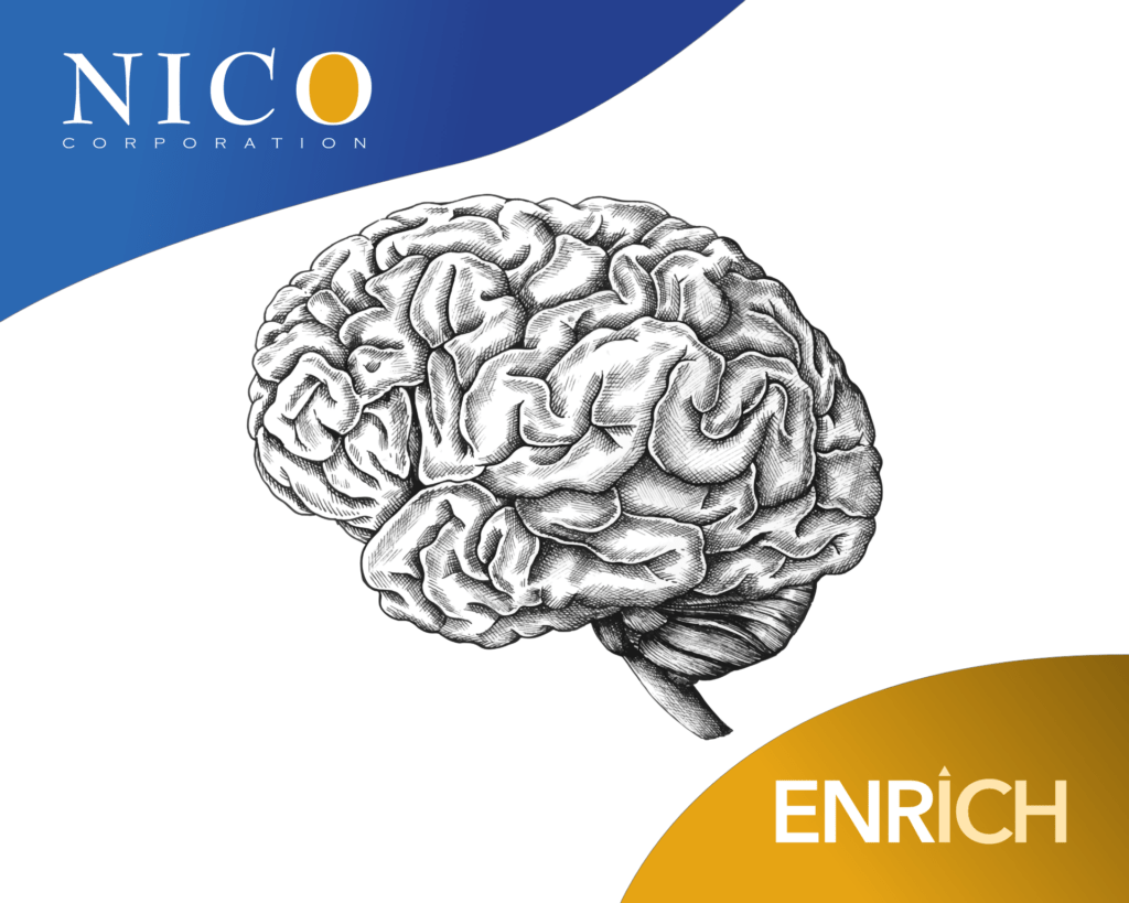
A hemorrhagic stroke is typically the most deadly and debilitating form of stroke, occurring in nearly two million people every year. Historically, it’s been treated by allowing the brain to bleed until hemorrhaging has subsided, leaving few recovery options for patients. About half of the individuals who suffer a hemorrhagic stroke die within 30 days.
But that could be changing—all thanks to a leading-edge stroke treatment that’s giving patients new hope.
After several years of the ENRICH (Early MiNimally-invasive Removal of ICH) trial, neurosurgeons were able to show positive results with improved outcomes for hemorrhagic stroke patients, and the results were shared at the 2023 American Association of Neurological Surgeons (AANS) annual meeting. The results were presented by representatives from Emory University and the NICO Corporation. Four current Goodman Campbell physicians, led by Dr. Charles G. Kulwin, participated in the trial. It involved 300 stroke patients at 37 treatment centers across the United States.
During the trial, the BrainPath device—a tool used to help surgeons get to the site of bleeding—carefully moved through the delicate folds and fibers of the brain. The BrainPath gently shifts the tissue to create a path to the site of the bleeding. Once there, the Myriad device, an automated suction and resection tool, can remove clotting.
These results were part of a stroke treatment trial in which surgeons hoped to improve the standard of care for hemorrhagic strokes beyond just waiting out the bleed.
“Goodman Campbell was one of the earliest sites and one of the largest contributors to the study,” said Dr. Kulwin. “Our hope is that this will finally take a difficult problem … and provide solid evidence that there is a correct way to manage it surgically.”
“Only a quarter of survivors get back to independence in the months following the stroke. A safe and effective way of operating on a hemorrhage by minimally invasive means has the potential to change the way we give care and save lives.”
We are proud to be part of this historical moment in stroke treatment, and we look forward to using this method to help save and improve the lives of our patients. We also look forward to continuing to find new, innovative ways to advance the field of neurosurgery. You can read more about the clinical trial here.
Goodman Campbell started way back in 1972, with the incorporation of the Indianapolis Neurosurgical Group established by Drs. Julius Goodman and John Russell.
Since then, we’ve pioneered new techniques in Indiana, such as the first microscopic discectomy, the first intracranial tumor embolization, and the first endoscopic placement of a VP shunt.
We’ve transformed ourselves by adding new disciplines—like interventional stroke care, minimally-invasive spinal surgery, interventional pain management, physical therapy, research, and education. We’ve steadily grown both our practice and our reputation, and we’ve never once looked back.
That is, until now.
This year, Goodman Campbell is turning 50 years old. Reaching that milestone has made us pause and reflect on our first half-century—and we could use your help documenting it.
If you’ve got photos, we’d love to see them. If you’ve got cards or notes, we’ll be glad to read them. If you have recordings, we’d be thrilled to hear them. If you’ve got videos, we can’t wait to watch them. Basically, if you’ve got anything at all that will help us expand our historical archives, we’ll be grateful for your assistance.
Visit our 50th Anniversary page now to submit your digitized photos or videos—or just to type in a favorite memory.
Don’t have access to a scanner? We’re still interested in what you have to share. Give us a call at 317.396.1300, and we’ll do what we can to make your Goodman Campbell memories last forever.
Until last August, Kelli McLaughlin of Carmel was living her life like normal. She was raising her four children with her husband, Ryan, volunteering for different causes, and managing two locations of her boutique shop, Clothes With a Cause, which donates proceeds to different charities throughout the year.
That month, Kelli started noticing odd, neurological symptoms and sensations in her body. She took a trip to the ER in early September and doctors didn’t find anything then. She returned later that month with her husband on their anniversary, because the sensations she was experiencing were very strong and interfering with normal tasks.
The sensations would be diagnosed as focal seizures, so brain scans were taken and they revealed Kelli had a tumor. Goodman Campbell neurosurgeon Dr. Troy Payner was assigned to Kelli’s case, and after close examination and tests, he delivered the news that she had glioblastoma, a rare brain cancer with no known cure. Ironically, Dr. Payner treated her father for several meningiomas 16 years ago.
“It took me about a month to wrap my head around my new situation. I was in complete disbelief because there were no warning signs whatsoever,” said Kelli. “But Dr. Payner and I instantly hit it off—I liked him when I met him years ago, and I like him now! My entire care team has been wonderful; they are great advocates and have helped me navigate every aspect of this process.”
Glioblastoma, a cancerous and incurable brain tumor, occurs in 3 out of 100,000 United States citizens annually. The median age of those diagnosed is 64.
Goodman Campbell treats roughly 50 cases each year. Symptoms and their severity vary with each person, but in general they include: headaches, seizures, nausea/vomiting, difficulty with speech, blurry vision, weakness on one side, confusion, or lethargy—more on signs, symptoms, and treatments here.
“Glioblastoma is the most common primary tumor to occur in the brain and it’s unfortunately cancerous. Despite tremendous research being done, we don’t have a cure. The goal of all treatments is to control the tumor as long as possible,” said Dr. Payner.
After the initial removal of the tumor, Kelli elected to start chemotherapy and radiation treatments, and wears an FDA-approved cap that helps slow tumor cells from replicating. Dr. Payner describes her as “an extremely ambitious woman who has an unflappable positive mental attitude.” Kathy Butz, a nurse on her care team, says that Kelli is very involved in her health care decisions, is open about her disease, and has a service-oriented heart.
“I don’t want to quit, I want to live. I’m committed to tackling this head-on, because I don’t see any other options,” Kelli said. “I feel like my purpose here is to make this place better than I found it, and help the next family that experiences something like this.”
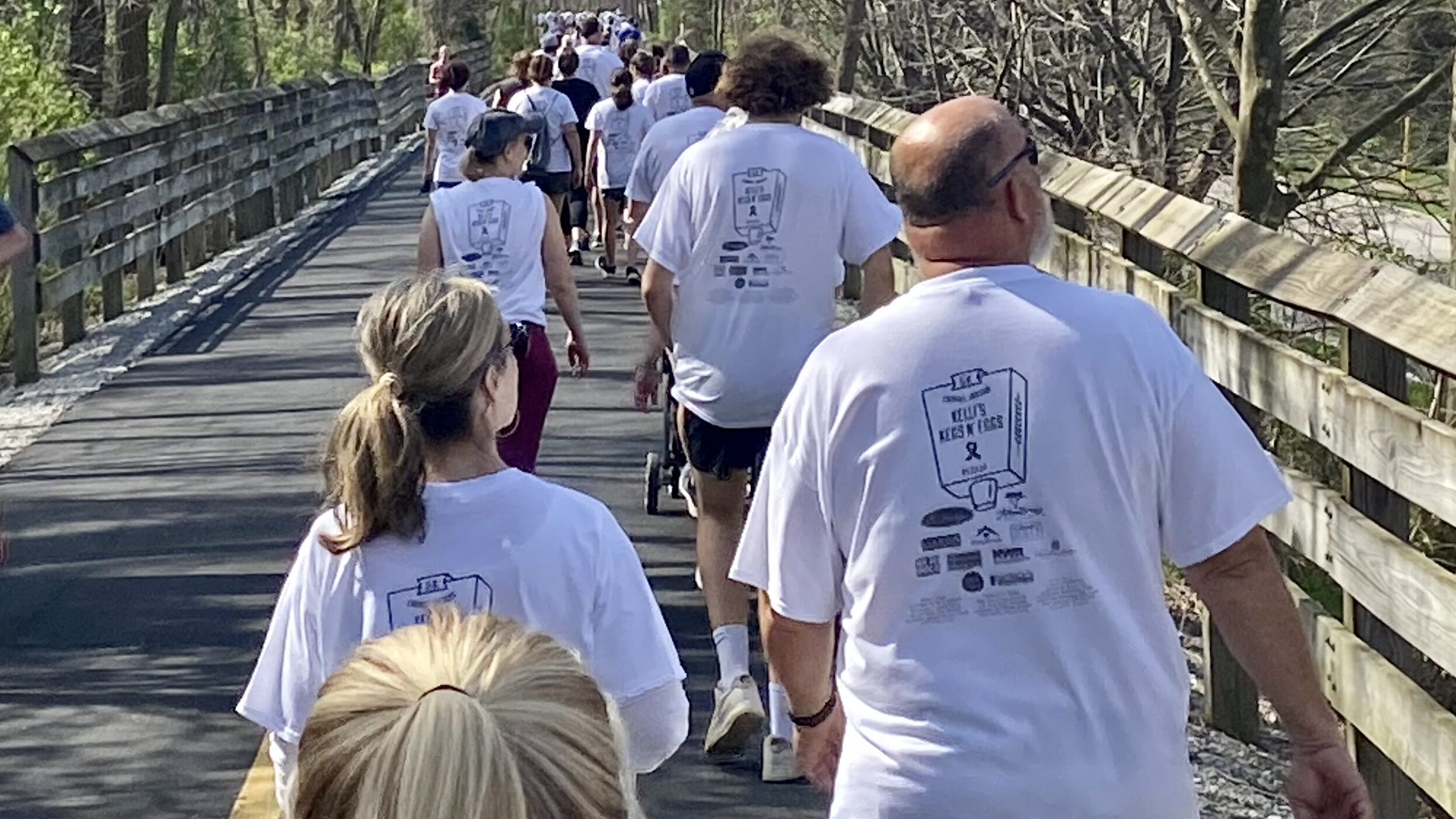
All of that shows in Kelli’s drive to help her community, despite her diagnosis and side effects from treatments. Along with her support network of friends and family, she recently organized the first “Kegs ‘N Eggs 5K” fundraising event. In addition to raising awareness for this disease, they successfully collected over $50,000 to help connect families going through a similar situation to available resources and treatments.
“I am so thankful for the great turnout and beautiful weather we had for the First Annual Kelli’s Kegs N Eggs 5K! Glioblastoma research is one of the most under-funded of all cancers, and it is my goal to change this,” said Kelli. “We want to raise awareness of this horrible disease and raise funds to ensure that other families do not have to suffer this terrible blow as we had to. It can be done and I firmly believe that there is a cure on the horizon.”

November 9, 2020
Jay and his family live in Indianapolis in a house that’s been in the family for several generations. Though his kids have grown, Jay looks back on a life in which sports and physical activity—whether on his own, with friends or with his wife and kids—have played a huge role.
Being active has been important not only physically and socially for Jay, but also mentally. When he’s not able get out and move regularly, it takes an emotional toll. “I need to be active, or I’m no fun to be around,” he said.
Because he’s been so active, he’s had his share of injuries; After battling back trouble on and off for years, Jay had gone six years with no significant problems when suddenly, while sitting on the sofa with his dog Agnes, his right leg went numb, became weak and wouldn’t wake back up.
He sought help from Dr. Mobasser at Goodman Campbell. “Dr. Mobasser sent me for an MRI and X-rays of my back to assess my spine,” Jay said. “It was a wreck. He said I needed to have this surgery, as I was developing nerve damage in my right leg. Dr. Mobasser made it clear that my condition was irreversible without surgery and would continue to get progressively worse. I was facing the rest of my life with a brace on my foot.”
Dr. Mobasser fused five vertebrae and freed up all the nerves being compressed at each of these levels. Having had some experience with knee surgeries, Jay was expecting a tough recovery, and Dr. Mobasser himself warned Jay that the recovery process from a multi-level lumbar spine surgery could be extremely difficult for the first few post-operative months.
His experience, however, was anything but difficult. “I feel incredibly lucky,” Jay said. “I never really had any pain. I sat right up and walked to a chair; I was walking the halls that same afternoon.”
He was in the hospital for less than two days. “The follow-up was great,” Jay said, “though it turned out I didn’t need it. They called once a week, just to make sure I was doing okay, and the next time I saw Dr. Mobasser was at my one-month checkup.”

“I’m back to living a normal life,” Jay said. “I’m biking, I’m walking, I’m playing with the dog. But you know, beyond that, it’s all the little things. Sleeping through the night. Getting dressed without having to contort myself. Even now, more than a year later, I’ll find myself doing something that used to be painful or difficult or impossible before the surgery, and it’s just such a relief to have that part of my life back. I credit Dr. Mobasser and Goodman Campbell with making that happen.”

The LITT procedure side effects most patients experience are temporary and manageable, especially when the procedure is performed by an experienced neurosurgical team. Because laser interstitial thermal therapy is minimally invasive and MRI guided, it is often associated with a shorter recovery and lower complication rates than traditional open brain surgery.
Still, any brain procedure deserves careful consideration. If you are researching laser interstitial thermal therapy risks, understanding potential complications and what recovery typically looks like can help you feel more informed and confident as you explore treatment options.
What Is the LITT Procedure?
Laser interstitial thermal therapy (LITT) is a minimally invasive, MRI-guided neurosurgical procedure that uses laser energy to precisely target and destroy abnormal brain tissue. It may be used for brain tumors, radiation necrosis, or epileptic foci.
During the procedure, a neurosurgeon creates a small incision and inserts a thin laser fiber into the targeted area of the brain. Real-time MRI temperature-mapping allows the surgeon to actively monitor heat distribution throughout the procedure, helping protect nearby healthy tissue. For many patients, LITT offers an alternative to traditional open craniotomy.
Who Is a Candidate for the LITT Procedure?
LITT may be appropriate for patients with certain brain tumors, including recurrent gliomas, metastatic tumors, or tumors located deep within the brain. It may also be used in patients with drug-resistant epilepsy. LITT radiation necrosis treatment may be appropriate following prior radiation therapy.
Not every patient is a candidate. Tumor size, location, overall health, and treatment goals all play an important role in determining whether LITT is appropriate. A detailed neurosurgical evaluation is essential.
What Are the Most Common Side Effects of LITT?
Most minimally invasive brain surgery side effects after LITT are usually temporary and improve as the brain heals.
Patients commonly experience:
- Post-surgical headaches caused by temporary swelling around the treatment area
- Temporary fatigue or grogginess after anesthesia
- Mild, temporary neurological changes, such as short-term memory difficulty, slight weakness, or word-finding challenges. These symptoms are often temporary and typically improve on their own over days or weeks.
Most patients stay in the hospital overnight and return home the next day.
How Long Do LITT Side Effects Typically Last?
Most aspects of LITT brain surgery recovery improve within days to a few weeks. Follow-up appointments are typically scheduled after treatment around the 1-month and 3-month marks so the neurosurgical team can monitor healing and recovery progress.
Regular follow-up imaging and neurological evaluations help ensure that concerns are addressed proactively.
What Are the Serious but Rare Risks of the LITT Procedure?
Although uncommon, MRI-guided laser ablation complications can occur, and we believe it’s important for patients to understand those risks clearly and honestly. Less common but more significant risks of the LITT procedure may include:
- Bleeding (hematoma) at the treatment site
- Infection, as with any surgical procedure
- Cerebrospinal fluid (CSF) leak
- Unintended thermal spread, meaning heat from the laser affects nearby healthy tissue
- Rare permanent neurological changes, such as weakness, speech changes, or vision effects (in the majority of cases, these improve over time)
Importantly, real-time MRI guidance and continuous temperature monitoring are specifically designed to minimize unintended thermal spread and help protect healthy brain tissue throughout the procedure. These are not passive safety measures. They are active tools the neurosurgeon uses in real time to guide treatment with precision.
While every surgical procedure carries some degree of risk, complication rates associated with LITT are generally lower than those seen with traditional open craniotomy procedures, particularly because LITT is minimally invasive and requires a much smaller surgical opening.
Why Choose Goodman Campbell for the LITT Procedure?
At Goodman Campbell Brain and Spine, we are proud to be a global leader in brain and spine care and a respected center for neurosurgery training and clinical research in the United States.
Consistently regarded as among the best in the nation, we make it easier for more people to access truly specialized care from leaders in neurosurgery and related specialties in the communities where we see patients. In addition, both patients and referring physicians have direct access to our team throughout the treatment process.
To discuss whether LITT may be an appropriate treatment for your condition, schedule a consultation today.
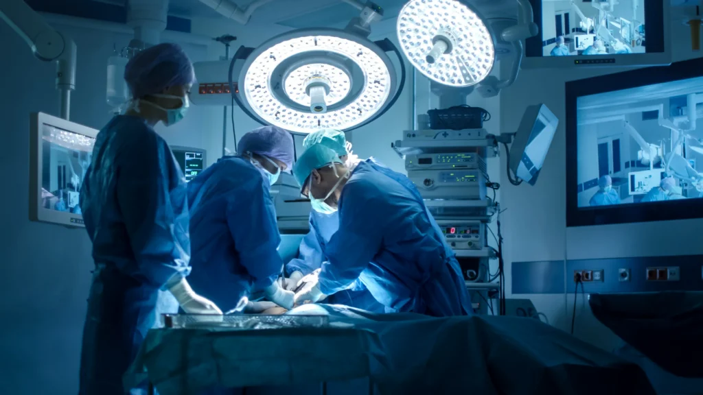
Persistent neck pain, arm pain, numbness, tingling, or weakness can make everyday activities difficult. When these symptoms are caused by a damaged disc or compression of the nerves or spinal cord, and nonsurgical treatment hasn’t provided adequate relief, ACDF surgery may be an option.
For some people, cervical spine symptoms can affect much more than comfort. Pain or weakness may interfere with work, sleep, exercise, or everyday tasks, while spinal cord compression can contribute to problems with balance, coordination, or hand function. When symptoms persist or neurological changes develop, identifying and addressing the underlying cause becomes particularly important. Over time, these symptoms may increasingly affect mobility, independence, and quality of life.
Surgery is not necessary for every cervical spine condition, but for appropriately selected patients, it may provide a way to relieve structural compression and protect neurological function.
Anterior Cervical Discectomy and Fusion (ACDF) is a commonly performed cervical spine procedure designed to address the structural source of nerve or spinal cord compression. If you are considering surgery, understanding how ACDF works, who may benefit, and what ACDF surgery recovery involves can help you make a more informed decision about your care.
What Is ACDF Surgery?
ACDF is a procedure used to remove a damaged or degenerative disc in the cervical spine, or neck, and relieve pressure on nearby nerves or the spinal cord.
During surgery, the neurosurgeon reaches the cervical spine through a small incision in the front of the neck. This anterior approach allows access to the affected disc without cutting through the larger muscle groups in the back of the neck or approaching the spinal cord from behind.
Once the damaged disc is removed, the neurosurgeon carefully removes the disc material or bone spurs causing compression. A bone graft and/or interbody spacer is then placed in the disc space to maintain proper spacing and alignment and promote fusion between the vertebrae. Additional hardware, such as a plate and screws, may be used to provide stability while the bones fuse.
Unlike treatments intended primarily to manage symptoms, ACDF is designed to address the underlying structural problem causing nerve or spinal cord compression. By removing the damaged disc and other material pressing on these structures, the procedure creates more space for the affected nerves or spinal cord. The fusion portion of ACDF then helps stabilize the treated area as the vertebrae heal together. For appropriately selected patients, this combination of decompression and stabilization can help address the source of symptoms such as radiating arm pain, numbness, tingling, or weakness, rather than focusing solely on managing those symptoms.
What Conditions Does ACDF Surgery Treat?
ACDF may be recommended for several conditions affecting the cervical spine, including:
- Cervical disc herniation: A damaged disc can bulge or rupture, putting pressure on a nearby nerve or the spinal cord.
- Degenerative disc disease: Age-related changes can cause cervical discs to lose height or deteriorate, potentially contributing to instability or compression.
- Cervical radiculopathy: A compressed nerve root can cause pain, numbness, tingling, or weakness that travels from the neck into the shoulder, arm, or hand. When nonsurgical cervical radiculopathy treatment is unsuccessful, surgery may be considered.
- Cervical myelopathy: Compression of the spinal cord can affect balance, coordination, walking, hand function, and other neurological abilities. Cervical myelopathy surgery may be recommended to relieve this pressure and help prevent further neurological decline.
- Bone spurs: Arthritic changes can cause extra bone to develop around the vertebrae and joints, narrowing the space available for nerves or the spinal cord.
When structural compression is present, ACDF can physically remove the source of that compression. Interventional pain techniques may help manage certain symptoms in appropriately selected patients, but they do not remove spinal cord compression or treat cervical myelopathy.
Is ACDF Surgery the Same as Other Cervical Spine Surgeries?
ACDF is one of several procedures neurosurgeons can use to treat cervical spine conditions.
Posterior cervical decompression and fusion approaches the cervical spine from the back of the neck and may be appropriate when compression, instability, or disease involves multiple areas. A minimally invasive posterior discectomy also approaches from the back and can relieve certain types of nerve compression without fusion.
Another option is artificial cervical disc replacement. While ACDF removes the damaged disc and fuses the surrounding vertebrae, artificial disc replacement uses an implant designed to preserve motion at the treated level. When comparing ACDF vs. disc replacement, the appropriate choice depends on factors such as the location and extent of the damage, the number of affected levels, spinal stability, and the patient’s individual anatomy.
There is no single best procedure for every patient. The location of compression, number of affected levels, spinal alignment, overall health, and other factors all influence which approach may be appropriate. At Goodman Campbell, our neurosurgeons perform multiple cervical spine approaches, allowing us to recommend the procedure that is best suited to each patient’s specific anatomy and condition. Rather than taking a one-size-fits-all approach, we evaluate the underlying cause and location of the problem, among other factors, to determine which surgical option may provide the most appropriate path forward.
Who Is a Candidate for ACDF Surgery?
ACDF surgery candidates typically have a cervical spine condition that is causing persistent symptoms or neurological changes and can be clearly identified through imaging and a neurological evaluation.
Surgery may be considered when:
- Neck or radiating arm symptoms continue despite an appropriate course of nonsurgical treatment
- Physical therapy or physical therapy-based rehabilitation has not provided adequate improvement
- Interventional pain management has not provided sufficient symptom relief for conditions appropriately treated with these approaches
- Weakness, numbness, or other neurological symptoms are progressing
- Imaging shows a herniated disc, bone spur, or another structural problem compressing a nerve or the spinal cord
- Imaging and symptoms indicate spinal cord compression or cervical myelopathy
You can learn more about the different surgical approaches in our Guide to Spine Surgery for a Cervical Herniated Disc.
Ultimately, determining candidacy requires an individualized evaluation. A neurosurgeon can review your symptoms, imaging, health history, and response to previous treatment before recommending the appropriate next step.
When Is It Time to Consider Surgery Over Conservative Treatment?
Many cervical spine conditions are initially treated without surgery. When symptoms are manageable and there is no significant or progressive neurological impairment, nonsurgical care may provide adequate relief.
Surgery becomes a more important consideration when symptoms continue despite appropriate conservative care or begin to interfere substantially with everyday life. Progressive neurological changes, including worsening weakness, numbness, hand coordination problems, or difficulty with balance or walking, can also indicate a need for prompt evaluation.
Imaging is another important part of this decision. If scans show a structural abnormality that corresponds with your symptoms, surgery may provide a way to directly address the source of the problem.
This distinction is particularly important when spinal cord compression is present. Spinal cord compression surgery is designed to physically relieve pressure on the spinal cord. The goal is not simply to mask symptoms, but also to address the structural cause and, when possible, protect neurological function.
What Happens During ACDF Surgery?
Although every surgical plan is individualized, the basic ACDF procedure steps generally include:
- Anesthesia: ACDF is performed under general anesthesia.
- Anterior incision: The neurosurgeon makes a small incision in the front of the neck.
- Accessing the cervical spine: Structures in the neck are carefully moved aside to provide access to the affected vertebrae and disc.
- Removing the damaged disc: The neurosurgeon removes the damaged disc to create access to the areas requiring decompression.
- Decompression: Disc material, bone spurs, or other tissue pressing on the nerve roots or spinal cord are carefully removed to relieve compression.
- Restoring the disc space: A bone graft and/or interbody spacer is placed in the space where the disc was removed to maintain disc height and alignment and provide a setting for fusion.
- Stabilization: Depending on the surgical technique and implant used, a plate and screws or other fixation may be used to stabilize the treated segment while fusion occurs.
- Closing the incision: Once the reconstruction is complete, the incision is closed.
You can find additional information about the procedure on our Anterior Cervical Decompression and Fusion procedure page.
ACDF single and multilevel surgeries follow the same general principles. A single-level procedure treats one disc space, while multilevel ACDF addresses two or more. The number of levels involved can affect surgical time, recovery, and the overall treatment plan.
Most ACDF procedures can be performed on an outpatient basis, allowing patients to return home the same day. Individual circumstances may require an overnight stay or additional observation.
How Long Does ACDF Surgery Take?
The answer depends on the number of levels being treated, the complexity of the compression, the patient’s anatomy, and whether additional reconstruction is necessary.
Many ACDF procedures take approximately 1 to 3 hours, with single-level procedures generally requiring less time than more complex multilevel surgeries.
Keep in mind that the time spent in the operating room is only part of your surgical day. Preoperative preparation, anesthesia, and postoperative monitoring in the recovery area add additional time. Your neurosurgeon can give you a more individualized estimate based on your planned procedure.
What Does ACDF Surgery Recovery Look Like?
Your ACDF recovery timeline begins immediately after surgery. Most patients are able to return home the same day, although this varies according to the procedure and individual health needs.
During the first several days and weeks, some discomfort and stiffness around the neck and surgical site are expected. Temporary swallowing difficulty or throat irritation can also occur because of the anterior surgical approach.
Your care team will provide specific instructions about activity, neck positioning, lifting, driving, incision care, and other restrictions. Walking and gradually increasing activity are often important parts of early ACDF surgery recovery, but it is essential to follow the recommendations provided by your surgical team rather than advancing activity too quickly.
Return to light or desk-based work varies. Some patients may return within approximately 2 to 6 weeks, depending on how well they are recovering and the physical demands of their job. More strenuous work may require additional time.
Physical therapy, when needed, may begin around 6 weeks after surgery or at another point recommended by your neurosurgeon. Not every patient will require the same rehabilitation plan.
It is also important to distinguish feeling better from being fully healed. The fusion process continues long after the incision heals. Bone fusion may take approximately 3 to 12 months to complete.
Your personal recovery may differ based on your age, overall health, the number of levels treated, the complexity of surgery, and how closely you follow postoperative recommendations.
What Are the Risks and Possible Complications of ACDF Surgery?
Every surgical procedure carries potential risks. Understanding ACDF surgery risks can help you have a more informed conversation with your neurosurgeon and weigh the potential benefits against the possible complications.
Potential risks and side effects can include:
- Temporary difficulty swallowing, also called dysphagia
- Temporary hoarseness or voice changes
- Infection
- Rare nerve injury
- Incomplete fusion, or pseudoarthrosis
- Adjacent segment disease, in which the spinal levels above or below the fusion experience changes over time
Some patients also experience temporary neck, shoulder, or arm symptoms as tissues heal and previously compressed nerves recover.
ACDF has a long history of use in cervical spine surgery and a well-established safety profile when performed for appropriate indications. Your individual risks, however, depend on factors such as your overall health, diagnosis, anatomy, and the complexity of the procedure.
An experienced neurosurgical team can help you understand these risks in the context of your specific condition rather than relying solely on general statistics.
Why Does Surgeon Expertise Matter for ACDF Surgery?
ACDF takes place close to the spinal cord, nerve roots, major blood vessels, and other important structures in the neck. For that reason, the training and experience of the surgical team are extremely important.
When choosing a neurosurgeon for ACDF, consider not only whether a surgeon regularly performs the procedure, but also whether the team has experience managing the full spectrum of cervical spine disease. Both simple and complicated cases require the same careful understanding of spinal anatomy, neurological function, imaging, and surgical decision-making.
At Goodman Campbell, our neurosurgeons treat both straightforward and highly complex spine conditions, allowing patients to receive specialized care even when their diagnosis or anatomy requires a more advanced approach.
Patients and referring physicians also have direct access to their Goodman Campbell team. Prompt appointment availability can help patients move efficiently from evaluation toward answers and, when appropriate, treatment.
What Sets A Neurosurgeon Apart for Cervical Spine Surgery?
Neurosurgery training is particularly extensive and specialized, with deep emphasis on the brain, spinal cord, nerve roots, and other structures of the nervous system. This training provides an important foundation for procedures such as ACDF, where relieving pressure on delicate neurological structures is a central goal of surgery.
Orthopedic surgeons also receive surgical training involving the musculoskeletal system and may pursue additional spine specialization. Neurosurgical training, however, involves more extensive and specialized training focused specifically on the nervous system, including the spinal cord and nerve structures involved in cervical spine surgery.
Goodman Campbell is a global leader in neurosurgery, neurosurgical training and clinical research. Our experience extends across the spectrum of brain and spine conditions, allowing us to bring that depth of expertise to each patient’s evaluation and surgical plan.
Are You a Potential Candidate for ACDF Surgery?
Living with persistent neck or arm pain, numbness, tingling, weakness, or other neurological symptoms can leave you wondering whether surgery is the appropriate next step. The answer depends on much more than a single symptom or imaging result.
At Goodman Campbell, we evaluate each patient individually, taking into account your diagnosis, symptoms, imaging, overall health, and previous treatment. Whether your condition is straightforward or highly complex, you have access to a globally recognized neurosurgery team with extensive experience treating disorders of the spine and nervous system. Efficient scheduling helps you move from uncertainty toward answers and an individualized treatment plan.
For most patients, the first step is not necessarily deciding whether to have surgery, but rather, understanding what is causing their symptoms and which treatment options make sense for their specific condition. A thorough evaluation can help determine whether ACDF, another cervical spine procedure, or a nonsurgical approach may be appropriate, while giving you the opportunity to ask questions about potential benefits, risks, recovery, and what you can realistically expect from treatment.
If you are experiencing cervical spine symptoms, we can help you understand your diagnosis and treatment options. Call 317-396-1300 to speak with our team or request an appointment online.
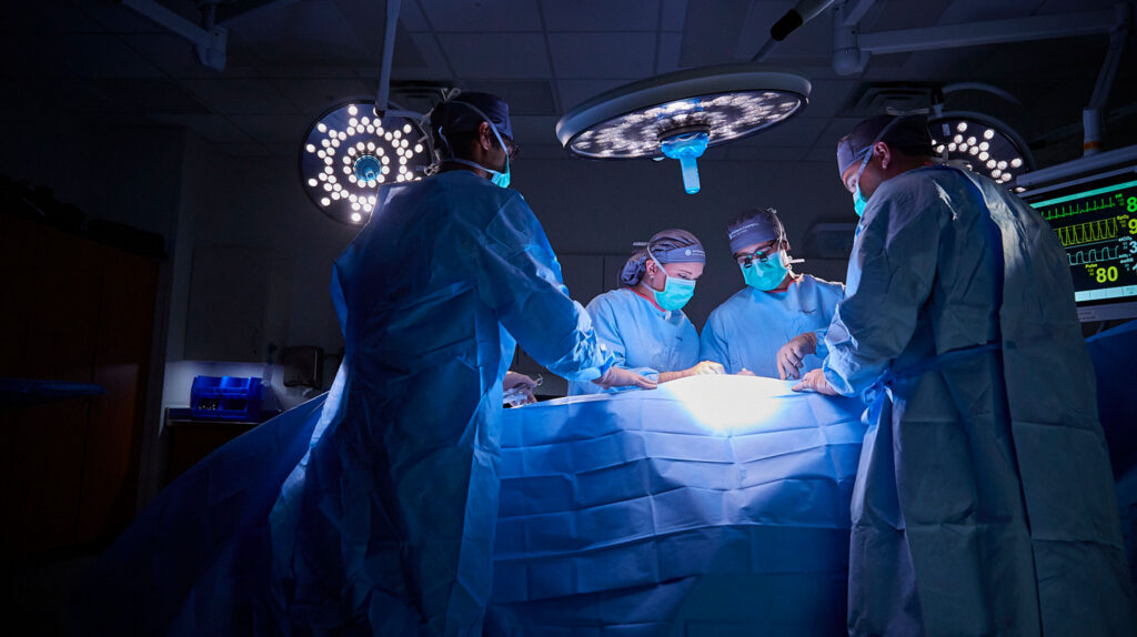
If you are considering Anterior Cervical Discectomy and Fusion (ACDF), you probably have some questions about what to expect, what to consider, and how major the surgery actually is. Let’s dive in.
Is ACDF Considered Major Surgery?
Yes, ACDF is considered major surgery. The procedure is performed near the spinal cord and nerve roots, requires general anesthesia, and involves a period of recovery afterward.
But “major surgery” does not necessarily mean unusually dangerous surgery. ACDF is one of the most commonly performed and well-studied spine procedures, with a strong safety record when performed by an experienced neurosurgical team. In fact, most patients undergoing ACDF are able to return home the same day.
ACDF is designed to address the structural source of symptoms. During the procedure, your surgeon removes a damaged disc that is compressing a nerve root or the spinal cord, and then stabilizes the affected area of the cervical spine through fusion.
What Are the Real Risks of ACDF Surgery?
It is reasonable to have questions about ACDF surgery risks, particularly because the procedure takes place close to the spinal cord and nerves.
As with any major procedure, anterior cervical discectomy and fusion complications are possible. Temporary difficulty swallowing, or dysphagia, is the most common early side effect. Some patients may also experience temporary hoarseness. Other potential cervical spine surgery risks include:
- Infection
- Bleeding
- Rare nerve injury
- Incomplete bone fusion, which may require future intervention
Serious complications are uncommon. The surgical approach — which involves the surgeon accessing the cervical spine through the front of the neck — is designed to reach the affected disc without disturbing the spinal cord or major muscle groups.
Your individual risk depends on several factors, including your overall health and the specifics of your cervical spine condition. Your surgical team can discuss these considerations with you before surgery so you understand the potential benefits and risks.
What Makes Surgeon Experience Important for Reducing ACDF Risks?
Because ACDF is performed close to the spinal cord, nerve roots, and surrounding vascular structures, your surgeon’s training and experience matter.
Neurosurgeons undergo extensive, specialized training focused on the brain, spine, spinal cord, and nervous system. This training is more extensive and specialized than orthopedic surgical training, particularly when it comes to operating around delicate neurological structures.
Choosing an experienced neurosurgeon for ACDF can help ensure that the complexities of your cervical spine condition are carefully evaluated and that your surgical plan accounts for the sensitive structures surrounding the treatment area.
What Does Recovery From ACDF Surgery Actually Look Like?
Recovery is an important consideration when thinking about undergoing ACDF. While individual timelines vary, the process generally involves gradually increasing activity while the neck and fusion heal.
Most patients can go home the same day or the day after surgery, depending on the extent of the procedure, their overall health, and how they are doing after surgery. During the first several weeks, patients typically have activity restrictions as the surgical site and neck heal. Physical therapy may begin around six weeks after surgery, although timing varies and some patients may not need formal physical therapy. Your surgeon will determine when rehabilitation is appropriate based on your progress.
Return to work also varies. Some patients with desk-based jobs may return to work within approximately 2 to 6 weeks, some with temporary restrictions. Jobs involving heavy lifting, repetitive neck movement, or other physical demands may require a longer recovery period. Although you may feel substantially better within weeks or months, the bone fusion itself takes considerably longer. Solid fusion can develop over several months and may take approximately 6 to 12 months to fully mature.
Your surgeon will monitor your recovery and provide individualized guidance about activity, work, and rehabilitation.
Is ACDF Surgery Worth It?
ACDF is a serious procedure in the sense that it requires preparation, anesthesia, and recovery. But for appropriately selected patients, it can help address the underlying structural cause of cervical nerve or spinal cord compression rather than simply managing symptoms.
The ACDF success rate and well-established safety profile can be reassuring for patients wondering, “Is ACDF surgery safe?” Your individual outcome, however, depends on your diagnosis, overall health, and other factors that your surgeon will evaluate.
For many patients, that balance is what makes ACDF worth considering. While surgery is never a decision to take lightly, treating the source of nerve or spinal cord compression may provide meaningful, lasting improvement in symptoms and function.
At Goodman Campbell, our globally recognized neurosurgery teams in Indiana and southeastern Wisconsin treat both straightforward and highly complex spine conditions. Our patients have direct access to their surgical team, and efficient scheduling can help you move seamlessly from evaluation toward answers and treatment — no separate referral needed.
If you are experiencing cervical spine symptoms, we can help you understand your diagnosis and treatment options. Call 317-396-1300 to speak with our team or request an appointment online.
By Vince Alentado, MD, Neurosurgeon, Spine Specialist
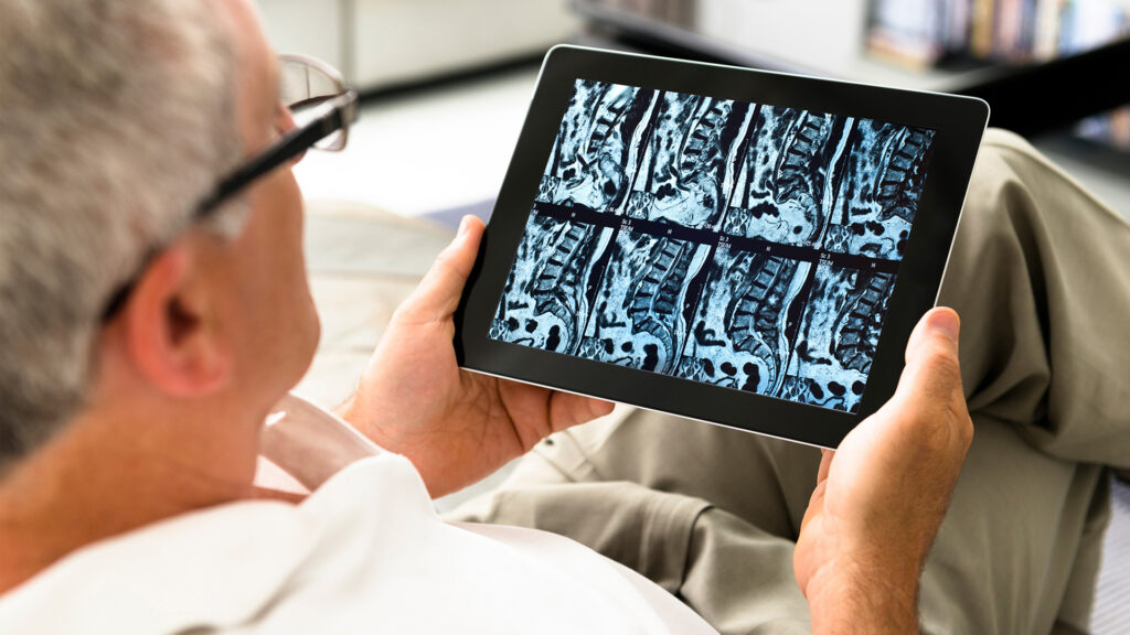
Around the time of the COVID-19 pandemic, patients gained unprecedented access to their medical records through electronic medical record patient portals. While this increased transparency and patient engagement is unquestionably valuable, it has also changed the way many patients approach their medical care.
Before widespread access to radiology reports, a patient with an MRI would typically come to the office with a constellation of symptoms, and the neurosurgeon would review the imaging in the context of the patient’s history and examination. Increasingly, patients began arriving to clinic having already read the radiology report — and often with significant anxiety about findings that may be incidental or clinically insignificant. I have seen dozens of patients who are asymptomatic but seek a neurosurgical consultation because of something concerning they read in an MRI report.
More recently, patients have taken this a step further by placing their radiology reports into their preferred artificial intelligence (AI) platform. As a result, patients are arriving with preconceived notions about what is happening with their spine, and they may also come with a list of AI-generated questions. I recently saw an 89-year-old patient who arrived with a detailed list of questions generated by ChatGPT.
AI undoubtedly has tremendous potential to improve healthcare, and patients should have meaningful access to their medical information. However, there is a risk that the abundance of information can inadvertently take some of the humanity out of the patient encounter. Instead of beginning with the patient’s symptoms, concerns, and examination and then using our neurosurgical expertise to interpret the imaging in that context, the conversation can become centered on what the MRI “shows.”
This is particularly important with degenerative spine imaging. Incidental degenerative findings are extremely common, even among healthy, asymptomatic individuals. Studies have demonstrated that by their 30s, approximately half of patients have evidence of disc degeneration, about one-third have disc height loss, and a substantial proportion have disc bulging. Radiology reports necessarily document many of these abnormalities, sometimes creating a lengthy list of findings that may have little or no clinical relevance to the patient’s symptoms. When an AI system then interprets a radiology report without the benefit of the patient’s history, physical examination, and clinical context, we can quickly arrive at a “garbage in, garbage out” scenario.
This expanding access to information places an additional burden on referring providers. Patients who become anxious after reading their radiology reports — or an AI-generated interpretation of those reports — may reach out to their primary care providers with questions and concerns before anyone has had the opportunity to put the findings into perspective.
For this reason, I believe it is increasingly important for referring providers to recognize the difference between common, benign radiographic findings and imaging abnormalities that warrant further evaluation. It is also worth considering early neurosurgical referral when advanced imaging is performed. Our role as neurosurgeons is not simply to interpret an MRI. It is to synthesize the patient’s history, symptoms, examination, imaging, and individual goals to determine what — if anything — needs to be done. In some cases, our most important contribution may be reassuring a patient that the frightening language in a radiology report does not represent a dangerous condition or a need for surgery.
As I tell my patients every day: We treat people, not pictures.
In an era of unprecedented access to medical information and increasingly sophisticated AI tools, I believe that principle is more important than ever. Rather than viewing AI as a replacement for clinical expertise, we should use it as a tool while ensuring that patients ultimately receive what an AI algorithm cannot provide: context, judgment, reassurance, and a human connection with their physician.
Learn more about neurosurgeon and spine specialist Vince Alentado, MD, who sees patients at Goodman Campbell – Noblesville. New patients can request an appointment online or call 317-396-1300.
You may be a good candidate for spinal cord stimulation if you have chronic nerve-related pain that hasn’t responded to conservative care and you’re open to a short trial period first.
If you’ve been living with chronic pain and searching for answers, you may have come across spinal cord stimulation (SCS) as a potential treatment option. However, many patients we meet are unsure whether they’re even a spinal cord stimulation candidate.
At Goodman Campbell, our goal is to make that clearer. Through our interventional pain management services, we help patients understand when this advanced option may be appropriate — and when it’s not. Our team combines the expertise of interventional pain management physicians and neurosurgeons to evaluate both simple and complex spine-related conditions.
What Is Spinal Cord Stimulation?
Spinal cord stimulation is a form of neuromodulation that works by interrupting pain signals before they reach the brain. A small device, called a spinal cord stimulator, delivers gentle electrical impulses to the nervous system, helping reduce the perception of pain.
Rather than masking symptoms, SCS targets how pain signals are processed. It’s typically considered only after more conservative treatments — such as physical therapy, nerve blocks, and injections — have not provided lasting pain relief. Explore a more comprehensive overview of spine care treatments at Goodman Campbell.
How Does an Interventional Pain Physician Evaluate Candidacy?
When you meet with an interventional pain physician at Goodman Campbell, we take a comprehensive look at your medical history, treatment history, and overall health. We evaluate your pattern of chronic pain symptoms, the type of persistent pain you’re experiencing, and how it affects your daily life.
A psychological evaluation is also part of the process, ensuring that a candidate for spinal cord stimulation is well positioned for success. These steps help us determine if we recommend spinal cord stimulation and whether it aligns with your goals for chronic pain relief.
Viable candidates for spinal cord stimulation can find themselves in a variety of circumstances before considering the procedure. Below are five common situations.
Sign 1: You’ve Exhausted Conservative Treatment Options
Spinal cord stimulation is not a first step. Most patients we see have already worked through more conservative treatments, including physical therapy and injection-based therapies (e.g., epidural steroid injections, nerve blocks, etc.)
If you’ve exhausted conservative treatment options without meaningful or lasting improvement, you may be a stronger spinal cord stimulation candidate. At Goodman Campbell, our team ensures that appropriate nonsurgical options have been fully explored before moving forward.
Sign 2: You Have Failed Back Surgery Syndrome or Persistent Post-Surgical Pain
Conditions like failed back surgery syndrome (FBSS) — persistent pain following spinal surgery — are among the most common reasons we recommend spinal cord stimulation.
Also known as post-laminectomy syndrome, this type of refractory pain often involves ongoing chronic back pain, sciatica, or leg pain after surgery. In many cases, additional surgery does not resolve the issue. Spinal cord stimulation offers a minimally invasive procedure that can provide meaningful pain relief without reopening the spine. When structural issues remain, Goodman Campbell may perform a microdiscectomy, but generally, SCS is utilized to address cases where further surgery isn’t the right next step.
Sign 3: Your Pain Is Neuropathic or Related to Complex Regional Pain Syndrome
Spinal cord stimulation is particularly effective for neuropathic pain, which originates from nerve damage.
This includes conditions such as:
- Peripheral neuropathy
- Sciatica
- Complex regional pain syndrome (CRPS)
These conditions often involve numbness, tingling sensations, and abnormal pain signaling. Because SCS works directly on those pain signals, it can be a powerful tool when other treatments fall short.
What Types of Conditions Does Spinal Cord Stimulation Treat?
At Goodman Campbell, we commonly use SCS for failed back surgery syndrome, CRPS, peripheral neuropathy, sciatica, peripheral neuropathy, and other forms of chronic pain that are not amenable to further surgical correction.
Spinal cord stimulation relieves chronic nerve-related pain; it does not open a narrowed spinal canal or correct structural compression. When stenosis is the root cause, we first evaluate whether decompressive treatment is appropriate.
Sign 4: Chronic Pain Is Significantly Limiting Your Daily Life
When persistent pain begins to control your life — affecting sleep, work, movement, or social activities — it may be time to consider a different approach.
The goal of spinal cord stimulation isn’t just to reduce discomfort. It’s also to restore function, improve quality of life, and help you get back to the activities that matter most to you.
Sign 5: You’re Willing to Undergo a Spinal Cord Stimulator Trial
One of the most unique aspects of SCS is the spinal cord stimulator trial.
Before any permanent implantation, patients undergo a 5-10 day trial period (or trial run) using temporary leads placed in the epidural space. This allows you to experience the potential benefits firsthand.
During the trial, the temporary leads connect to an external trial stimulator you wear outside the body, typically for about 5 to 10 days. We generally consider the trial a success when it delivers roughly 50% or greater pain reduction, along with meaningful gains in function and comfort. Because nothing is permanently implanted at this stage, the trial and the device are removable and reversible.
If the trial is successful — meaning you achieve meaningful pain relief — we move forward with permanent placement. If not, the leads are removed. This built-in evaluation step makes SCS a lower-risk option and a strong indicator of long-term success.
Who Is Not a Good Candidate for Spinal Cord Stimulation?
Spinal cord stimulation is not the right fit for everyone, and part of a caring evaluation is recognizing when it should be avoided.
You may not be a candidate for spinal cord stimulation if you have an active infection, an unmanaged bleeding disorder, or a health profile that makes you a poor surgical risk. Other reasons we may recommend a different path include an inability to operate the device, an unresolved significant psychological condition, pregnancy, or a trial that did not provide meaningful relief.
In these situations, we work with you to explore safer, more appropriate options.
| Good Candidate | Not a Candidate |
| Chronic nerve-related pain that hasn’t responded to conservative care | Active infection |
| Failed back surgery syndrome or persistent postsurgical pain | Unmanaged bleeding disorder |
| Neuropathic pain, CRPS, or peripheral neuropathy | Inability to operate the device |
| A trial with roughly 50% or greater pain reduction | Unresolved significant psychological condition |
| A cleared psychological evaluation | Poor surgical risk or pregnancy |
| Reasonable surgical risk and realistic goals | A trial that did not provide meaningful relief |
What to Expect After the Trial
If your trial provides meaningful relief, we move forward with permanent implantation. During this minimally invasive procedure, we place the permanent leads and a small generator, which is typically positioned just beneath the skin. Programming is tailored to your pain pattern, and the settings can be adjusted over time as your needs change.
In appropriately selected patients, published evidence shows that many people maintain meaningful, durable pain relief and improved function after permanent implantation, which is why the trial period is such an important step in choosing the right candidates.
Is a Consultation the Right Next Step for You?
If you’re wondering whether you might be a candidate for spinal cord stimulation, that curiosity is a great place to start. At Goodman Campbell, our interventional pain physicians work closely with our neurosurgeons to ensure every patient receives a thoughtful, individualized evaluation. Just as important, both patients and referring providers have direct access to their Goodman Campbell care team, making it easier to ask questions, stay informed, and feel confident every step of the way.
We’re also proud to be a national leader in SCS research, including active trials in neuromodulation and leading enrollment in the Abbott T2P study across seven national sites — bringing advanced pain management options directly to our patients.
If you’re ready to take the next step, call us or request an appointment here.
Frequently Asked Questions
How Do I Know if I’m a Candidate for Spinal Cord Stimulation?
You may be a spinal cord stimulator candidate if you have chronic nerve-related pain that hasn’t responded to conservative care, your pain significantly limits daily life, and you’re willing to try a short trial period first. The best way to know is through a comprehensive evaluation with our interventional pain physicians, which includes your medical and treatment history and a psychological evaluation. This is how we determine which patients are the best candidates for spinal cord stimulation.
Who Should Not Get a Spinal Cord Stimulator?
Spinal cord stimulation may not be appropriate if you have an active infection, an unmanaged bleeding disorder, an inability to operate the device, an unresolved significant psychological condition, poor surgical risk, or pregnancy, or if a trial did not provide meaningful relief. Understanding who is not a candidate for spinal cord stimulation helps us guide you toward safer, more effective options.
Does Spinal Cord Stimulation Treat Spinal Stenosis?
No. Spinal cord stimulation relieves chronic nerve-related pain; it does not open a narrowed spinal canal or correct structural compression. When stenosis is the root cause, we first evaluate whether decompressive treatment is appropriate.
What Happens During the Spinal Cord Stimulator Trial?
During the spinal cord stimulator trial, we place temporary leads in the epidural space and connect them to an external trial stimulator you wear outside the body. You go about your normal routine so you can experience the potential relief firsthand. If it delivers meaningful pain reduction, we move forward with permanent implantation; if not, the leads are simply removed.
How Long Does the Trial Last?
The trial typically lasts about 5 to 10 days. Success is generally judged by roughly 50% or greater pain reduction, along with meaningful improvement in function and comfort.
Can Spinal Cord Stimulation Help Peripheral Neuropathy?
Yes. A spinal cord stimulator for peripheral neuropathy can be an effective option, and the therapy is cleared for painful diabetic peripheral neuropathy. Because SCS works directly on how pain signals are processed, it can help when other treatments have fallen short.
Is Spinal Cord Stimulation Reversible?
Yes. Spinal cord stimulation is designed to be reversible. The trial device is fully removable, and even a permanent system can be turned off or removed if it no longer meets your needs.

If you’ve been living with chronic pain and still don’t have clear answers, you’re not alone. Many patients come to us after trying physical therapy or other treatments without lasting relief. That’s where interventional pain management and our interventional pain management services can make a real difference, not just by easing symptoms, but by helping identify the exact source of your pain.
At Goodman Campbell, nerve blocks are part of a precise, evidence-based diagnostic process. Our aim is to uncover the root cause of your pain, not merely mask it.
What Is a Nerve Block?
In plain terms, a nerve block is a small, targeted injection of numbing medication placed near a specific nerve. Nerve blocks can serve two important purposes: they can help identify the source of your pain and, when used therapeutically, provide targeted pain relief. When we use a nerve block to pinpoint where your pain is coming from, we call it a diagnostic nerve block. When the goal is to reduce pain and improve function, it is considered a therapeutic nerve block. Both approaches are valuable tools in interventional pain care and can help guide a treatment plan tailored to your needs.
How Does a Nerve Block Work?
The mechanism is simple. We place a local anesthetic right next to a specific nerve, and that medication temporarily numbs the nerve so it stops relaying pain signals to your brain. If your usual pain quiets down while the anesthetic is working, that nerve or the structure it serves is very likely the culprit. If nothing changes, we’ve learned something just as valuable and keep looking.
What Is a Diagnostic Nerve Block and How Is It Different From a Treatment?
A diagnostic nerve block is a targeted nerve block injection used as a diagnostic tool. Instead of simply providing pain relief, it serves as a diagnostic tool that helps your care team determine which nerve, joint, or spinal structure may be causing your symptoms.
In many cases, a spine surgeon may recommend a diagnostic nerve block when imaging studies and physical examination findings do not tell the full story. The information gained from the injection can help confirm whether a specific area is responsible for your pain and may play an important role in treatment planning, including determining whether surgery is likely to address your symptoms.
Using imaging guidance, an interventional pain management physician places a small amount of local anesthetic at a precise injection site, near a specific nerve or affected nerve. If your pain improves, it tells us that the nerve is likely responsible for sending those pain signals.
What Is the Difference Between a Diagnostic and a Therapeutic Nerve Block?
A therapeutic nerve block is designed to provide longer-lasting pain relief and help reduce inflammation.
A diagnostic nerve block, on the other hand, is about clarity. It helps answer questions such as:
- Which pain patterns are tied to which nerves?
- What is the underlying cause of your ongoing pain?
- Which particular nerve is responsible for sending those pain signals?
- Would treating a specific area be likely to improve your pain?
Often, both approaches work together as part of a larger treatment plan.
How Does a Nerve Block Help Find the Source of Your Pain?
Pain doesn’t always originate where you feel it. For example, nerve pain in the hip may actually stem from the spinal column. By temporarily interrupting pain messages along a particular nerve, we can see how your body responds. If your symptoms improve, we’ve likely identified the exact source.
This is exactly why a nerve block for back pain or stubborn hip discomfort can be so revealing. Pain felt in the hip or buttock often traces back to an irritated nerve root or facet joint in the spine, and a diagnostic block helps us confirm where it truly begins so we treat the real source rather than the symptom.
This is especially helpful in chronic back pain diagnosis, where multiple structures, such as discs, joints, or peripheral nerves, may be involved.
What Do the Results Mean if the Nerve Block Does or Doesn’t Work?
Your nerve block results guide the next steps:
- If you experience pain relief, we’ve identified the source and can focus treatment there.
- If you don’t experience pain relief, that nerve likely isn’t the cause, and we continue investigating.
Either way, the information moves your care forward.
We also look for an objective benchmark, not just a general sense of “better.” A positive diagnostic block is typically judged by a marked reduction in pain during the anesthetic’s window. Little or no relief redirects our search to another structure, which is just as important a finding.
What Types of Nerve Blocks Are Used to Diagnose Spine and Nerve Pain?
There are several types of nerve blocks we use in interventional spine care, including:
| Block Type | Structure It Targets/Evaluates | Primary Role |
| Medial branch block | Medial branch nerves that supply the facet joints | Primarily diagnostic; can also be therapeutic |
| Selective nerve root block | A specific spinal nerve root | Diagnostic (helps determine weather a specific nerve root is contributing to symptoms) |
| Sacroiliac (SI) joint block | The SI joint | Diagnostic or therapeutic |
| Epidural steroid injection | Epidural space and irritated nerve roots | Primarily therapeutic |
Each is chosen based on your symptoms and suspected pain conditions.
What Happens After a Diagnostic Nerve Block?
These procedures are performed on an outpatient basis, so you can go home the same day. We’ll ask you to track your symptoms closely. Specifically, we want to understand how much relief you feel, how long it lasts, and how it affects your daily activities. This feedback is critical in shaping your treatment options.
This information is an important part of the diagnostic process. Your response to the injection helps confirm whether a specific nerve, joint, or other spinal structure is responsible for your pain and helps guide the most appropriate treatment plan.
In some cases, we may recommend additional nerve blocks to confirm findings or further refine your spine pain diagnosis.
Is a Diagnostic Nerve Block Safe? What to Expect
Diagnostic nerve blocks are among the most established procedures in interventional pain care, and they’re considered very safe when performed by an experienced team. Because we use only a small amount of local anesthetic placed precisely under imaging guidance, side effects tend to be mild and temporary, and may include brief soreness at the injection site or short-lived numbness or weakness in the area while the anesthetic is active.
As with any injection, less common risks include minor bleeding, infection, or a reaction to the medication, which is one more reason it matters who performs your block and how carefully the site is prepared.
Here’s what to expect: the visit is quick and done on an outpatient basis, we numb the skin first, and you head home the same day. From there, your job is simply to notice how your body responds so we can turn that feedback into a clear plan. You can learn more about the full range of our spine care and how a diagnostic block fits into it.
Can a Nerve Block Lead to Surgery?
Yes. In some cases, diagnostic nerve blocks are used to help determine whether surgery is likely to address the source of a patient’s symptoms.
If a nerve block provides significant relief, it can help confirm that a specific nerve root, joint, or spinal structure is the pain generator. This information gives your care team greater confidence in recommending the most appropriate treatment, whether that involves continued conservative care, interventional pain management, or surgery.
Why Does It Matter Who Performs Your Nerve Block?
Precision matters.
Our interventional team has highly specialized training in the neck, spine, and nervous system, providing extensive, focused expertise. We understand how every structure in the spine connects and can contribute to pain.
We’re also proud to offer world-class neurosurgery expertise right here in Indiana, meaning that you don’t have to travel far for advanced care. As a global leader in back and spine care, we treat everything from straightforward cases to complex spine conditions. Our approach is backed by proven patient outcomes and efficacy data, and because you have direct access to our team, your care stays coordinated, clear, and focused on results.
If you’re ready to move beyond uncertainty and understand what’s truly causing your pain, Goodman Campbell is here to help. Request an appointment with our spine team to take the next step toward answers and lasting relief.
Frequently Asked Questions
Does a Nerve Block Cure the Cause of My Pain?
No. A diagnostic nerve block is a diagnostic tool, not a structural cure. It localizes and confirms the source of your pain and can offer temporary relief while the local anesthetic is active, but it doesn’t remove structural compression like a disc pressing on a nerve root.
Can Hip Pain Actually Come From My Spine?
Yes. Pain doesn’t always originate where you feel it, and hip or buttock pain frequently stems from an irritated nerve root or facet joint in the lower spine. A diagnostic nerve block helps us tell true hip-joint pain from spine-referred pain so we can treat the real source.
Is a Diagnostic Nerve Block Painful?
Most patients tolerate the procedure well. We numb the skin first, use imaging guidance for precise placement, and the injection itself is brief. You may feel a quick pinch or some pressure. It’s an outpatient procedure, and you go home the same day.
How Soon Will I Know My Results?
You’ll often notice how you respond within the first hours, while the local anesthetic is active. We ask you to track how much relief you feel, how long it lasts, and how it affects your daily activities, and then we review that feedback together to plan your next step.

If you’ve been dealing with neck pain for more than a few days, it’s natural to wonder: Is neck pain serious? The good news is that many cases are caused by muscle strain, poor posture, or overuse and improve with time. However, some symptoms can signal a more significant problem involving the cervical spine.
Understanding the difference between routine discomfort and serious neck pain can help you decide when to see a doctor for neck pain and whether your symptoms require specialist evaluation.
When Is Neck Pain Just Muscle Strain – and When Is It Something More?
Many people struggle to determine when they’re suffering from a stiff neck vs. nerve pain. In many cases, everyday neck pain is caused by muscle strain related to posture, sleeping position, tension, or overuse. This type of pain is typically limited to the neck, feels sore or stiff, and gradually improves on its own over days or weeks.
Structural cervical spine conditions are different. Rather than involving muscles alone, these problems affect the discs, nerves, or spinal cord within the neck. When a nerve root becomes compressed, symptoms often extend beyond the neck and into the shoulder, arm, or hand.
When the spinal cord becomes compressed, symptoms may include hand weakness or difficulty with coordination and fine motor tasks, as well as problems with balance, walking, and even leg function.
Understanding this distinction is important. Muscle-related neck pain is often self-resolving. Structural conditions involving nerve compression or spinal cord involvement are less likely to improve without targeted treatment and often require specialist evaluation.
How Long Should Neck Pain Last Before You See a Doctor?
One of the most common concerns we hear is about neck pain that won’t go away. If symptoms persist for more than a few weeks, continue to worsen, or interfere with daily activities, it may be time for a medical evaluation.
You should seek care sooner if you experience pain radiating into the arm, weakness, numbness, tingling, or changes in coordination.
What Are the Warning Signs That Neck Pain Is Serious?
Recognizing neck pain red flags can help you determine whether your symptoms may be related to a structural cervical spine problem.
One of the most common serious neck pain symptoms is neck pain radiating to the arm, shoulder, or hand. This may indicate cervical radiculopathy, a condition caused by compression of a nerve root in the neck.
Common cervical radiculopathy symptoms include arm pain, burning sensations, weakness, and tingling that travels into the hand or fingers.
If you begin dropping objects, struggle with hand coordination, or notice increasing arm weakness, the problem may involve more than a single nerve. These symptoms may indicate ongoing nerve compression and should not be ignored.
Particularly concerning are cervical myelopathy warning signs such as difficulty walking, balance changes, leg weakness, clumsiness, or loss of coordination. These symptoms may indicate cervical myelopathy caused by spinal cord compression and require prompt neurosurgical evaluation.
Neck pain following a fall, accident, or other trauma should also be evaluated, as it may indicate a fracture or ligament injury.
Finally, severe neck stiffness accompanied by fever and a severe headache could indicate meningitis and requires emergency medical care.
What Does It Mean When Neck Pain Radiates Down Your Arm?
When neck pain begins to radiate to your arm, it often means a nerve is being irritated or compressed. Common causes include a herniated disc or age-related changes that narrow the spaces around the nerves.
Because nerves control both sensation and muscle function, compression can cause pain, numbness, tingling, weakness, or a combination of symptoms. Treating the underlying cause is often more effective than simply addressing symptoms.
When Does Neck Pain Require a Neurosurgeon?
Neck pain may warrant neurosurgical evaluation when symptoms or imaging suggest nerve root compression, spinal cord compression, cervical myelopathy, a herniated disc, or another structural cervical spine condition.
Interventional pain physicians can play an important role in helping patients manage certain chronic neck pain conditions that do not involve significant structural compression. However, structural problems affecting the spinal cord or nerve roots require a different kind of expertise. Conditions such as spinal cord compression and cervical myelopathy require neurosurgical evaluation because the goal is not symptom management but rather addressing the underlying cause of neurological dysfunction.
At Goodman Campbell, our neurosurgeons are trained to treat both simple and complex cervical spine conditions, including many that extend beyond the scope of orthopedic specialists or general practitioners. We evaluate the entire cervical spine, identify the root cause of symptoms, and develop treatment plans focused on long-term function and quality of life.
Many patients do not require surgery. Depending on the diagnosis, treatment may include physical therapy and other conservative approaches, including interventional pain management. When surgery is appropriate, our team offers advanced cervical spine procedures, including anterior cervical discetomy and fusion (ACDF), cervical disc arthroplasty, and minimally invasive options, when appropriate.
Our patients benefit from having access to a world-class neurosurgery team right here in Indiana. We provide direct access to our care team and treatment supported by extensive patient outcomes data.
If you’re concerned about ongoing neck pain, arm symptoms, or possible signs of nerve or spinal cord compression, request an appointment directly online.

Severe Neck Pain: What Are the Most Common Causes
Severe neck pain can be alarming, especially when it appears suddenly, persists for weeks, or interferes with everyday activities. While many cases of neck discomfort are caused by temporary muscle strain, severe pain may sometimes indicate an underlying condition affecting the spine, discs, nerves, or surrounding structures. Understanding the most common causes of severe neck pain can help you recognize when symptoms may require professional evaluation and treatment.
What Makes Neck Pain “Severe” and Why It Matters
Most people experience neck pain from time to time. A poor sleeping position, a long day at a desk, or an awkward movement can leave your neck feeling stiff and sore for a few days.
However, some severe neck pain causes are very different from ordinary muscle strain.
When pain becomes intense, persistent, progressively worse, or accompanied by neurological symptoms, it may indicate a structural problem involving the discs, nerves, joints, or spinal cord.
Many patients with severe neck pain wonder whether they’re dealing with a simple strain or something more serious. Understanding the difference is important because structural conditions often require a different evaluation and treatment approach than temporary muscle soreness.
How Do You Know if Your Neck Pain Is a Warning Sign?
For many people, neck pain is caused by strained muscles, irritated soft tissues, or temporary inflammation. This type of discomfort often improves within a few days or weeks and tends to stay localized to the neck and upper shoulders.
Structural problems are different. When a disc, nerve, or the spinal cord becomes involved, symptoms often become more persistent, more severe, or begin affecting other parts of the body. Structural problems are more likely when symptoms persist, recur frequently, or are accompanied by numbness, weakness, balance changes, or pain that travels beyond the neck.
While these symptoms do not always indicate a serious condition, they may suggest that the problem extends beyond ordinary muscle soreness and warrants further evaluation. The longer neurological symptoms continue, the more important it becomes to identify the underlying cause and determine whether nerve or spinal root compression is present.
What Are the Red Flags That Require Immediate Evaluation?
Some neck pain red flags should never be ignored, including:
- Significant arm or hand weakness
- Difficulty walking or maintaining balance
- Loss of coordination or grip strength
- Changes in bowel or bladder function
- Persistent severe neck stiffness combined with worsening neurological changes
These symptoms may indicate spinal cord involvement and should be evaluated promptly.
What Are the Most Common Structural Causes of Severe Neck Pain?
Several conditions can cause significant neck pain, and the correct diagnosis depends on identifying the underlying structural problem.
Cervical Herniated Disc
A herniated disc occurs when one of the discs between the vertebrae in the neck bulges or ruptures, placing pressure on a nearby nerve. This is one of the most common causes of severe neck pain and may also cause pain, numbness, tingling, or weakness that travels into the shoulder, arm, or hand.
Cervical Spinal Stenosis
Cervical spinal stenosis refers to a narrowing of the spaces within the spinal canal. As that space becomes smaller, nerves or the spinal cord can become compressed. Stenosis often develops gradually as part of the aging process and may cause neck pain, arm symptoms, balance problems, or walking difficulties.
Cervical Spondylosis
Cervical spondylosis is the age-related wear and tear that affects the discs, joints, and bones of the neck. It can lead to stiffness, chronic neck pain, bone spur formation, and narrowing around the nerves or spinal cord.
Cervical Myelopathy
Cervical myelopathy occurs when the spinal cord itself becomes compressed. Because the spinal cord carries signals between the brain and the rest of the body, compression can cause symptoms beyond neck pain, including difficulty walking, balance problems, loss of hand coordination, weakness, and changes in bowel or bladder function. Cervical myelopathy is generally considered more serious than nerve root compression alone and requires evaluation by a neurosurgeon.
Cervical Radiculopathy
Cervical radiculopathy occurs when a cervical nerve root becomes compressed or irritated. Unlike cervical myelopathy, which affects the spinal cord, radiculopathy affects an individual nerve as it exits the spine. Symptoms commonly include neck pain radiating into the shoulder or arm, numbness, tingling, burning sensations, or weakness along the path of the affected nerve.
Is a Herniated Disc Always the Cause of Radiating Neck Pain?
A herniated disc is a common cause of neck pain and arm symptoms, but it is not the only possible explanation for pain that radiates beyond the neck.
When a cervical disc herniates and compresses a nearby nerve root, it can cause neck pain, numbness, tingling, weakness, or neck pain radiating to the arm. However, similar symptoms can also occur when nerve roots are compressed by bone spurs, cervical spinal stenosis, or other degenerative changes within the cervical spine.
Several structural conditions can produce similar symptoms, and each may require a different treatment approach. The key is determining exactly what is causing the nerve compression so that treatment addresses the underlying condition rather than simply managing symptoms.
What Is the Difference Between Cervical Stenosis and Cervical Myelopathy?
Cervical spinal stenosis refers to narrowing within the spinal canal. Common cervical spinal stenosis causes include age-related degeneration, disc changes, bone spurs, and thickened ligaments.
Not everyone with stenosis develops symptoms. However, when narrowing begins to compress the spinal cord itself, the condition may progress to cervical myelopathy.
Cervical myelopathy occurs when the spinal cord becomes compressed. Unlike nerve root compression alone, spinal cord compression can affect balance, coordination, walking ability, hand function, and even bowel or bladder control.
What Symptoms Indicate the Cause Is More Than Muscle Pain?
Muscle soreness usually remains localized to the neck and upper shoulders. Structural conditions often produce symptoms that extend beyond the painful area. Common signs of nerve involvement include:
- Pain radiating into the shoulder or arm
This often suggests irritation or compression of a cervical nerve root rather than a simple muscle strain. - Numbness or tingling in the fingers
Sensations such as “pins and needles” may occur when nerve signals are disrupted by pressure within the cervical spine. - Weakness in the arm or hand
Difficulty lifting objects, opening jars, or performing routine tasks can indicate that nerve function is being affected. - Difficulty walking or balance changes
Problems with balance, coordination, or walking may indicate that pressure is affecting the spinal cord rather than a single nerve root.
- Difficulty gripping objects
Reduced hand coordination or strength may be a sign that nerve compression is progressing. - Bowel/bladder changes
New difficulty controlling bowel or bladder function is uncommon, but it can be a serious sign of spinal cord compression and should be evaluated promptly.
When Does Neck Pain Mean Something Is Wrong With the Spinal Cord?
Certain symptoms may indicate spinal cord compression rather than simple nerve irritation. These include difficulty walking, balance problems, loss of hand coordination, frequent dropping of objects, weakness in the arms or hands, or changes in bowel or bladder function.
While these symptoms do not automatically mean surgery is necessary, they do warrant prompt evaluation by a spine specialist trained to diagnose and treat spinal cord compression.
How Is the Cause of Severe Neck Pain Diagnosed?
Accurate diagnosis begins with a detailed medical history and physical examination. We evaluate symptoms, neurological function, strength, reflexes, coordination, and balance to better understand the source of the problem.
Imaging studies may also be recommended, including:
- X-rays to evaluate alignment and degenerative changes
- MRI scans to assess discs, nerves, and the spinal cord
- CT scans to provide additional detail about bony structures
The goal is not simply to identify where pain is occurring, but to determine what is causing it. That distinction helps guide the most appropriate treatment plan.
Why Does It Matter Whether a Neurosurgeon or Orthopedic Surgeon Evaluates My Neck?
Both neurosurgeons and orthopedic spine surgeons treat cervical spine conditions, including herniated discs, spinal stenosis, nerve root compression, and cervical myelopathy. However, when symptoms involve the spinal cord or nerve roots, a neurosurgeon’s specialized training in the diagnosis and surgical treatment of disorders affecting the nervous system can be particularly valuable.
Conditions such as cervical myelopathy, spinal cord compression, and complex nerve root compression require careful evaluation of both the spinal structures and their effect on neurological function. A neurosurgical evaluation can help determine whether symptoms are coming from the spinal cord, nerve roots, or another structural abnormality and whether surgery may be appropriate.
When Should You See a Neurosurgeon for Severe Neck Pain?
If your symptoms are worsening, radiating into your arms, causing weakness, affecting your balance, or limiting your daily activities, it may be time to seek specialist care.
At Goodman Campbell, our neurosurgeons are trained to treat both simple and complex cervical spine conditions, a level of specialization that extends beyond traditional orthopedic spine training. As a global leader in neurosurgery with access to interventional pain management physicians as well, we provide patients access to one of the most accomplished neurosurgical teams in the world, right here in Indiana.
Our team works closely with physical therapists and other specialists to identify the root structural cause of symptoms and develop an individualized treatment plan. When surgery is necessary, we offer advanced procedures, including microdiscectomy and other proven surgical techniques, supported by extensive patient outcomes data.
If you are wondering when to see a doctor for neck pain, especially when symptoms involve nerve compression, arm weakness, balance changes, or possible spinal cord involvement, early evaluation can help prevent further progression.
Learn more about cervical spine treatment options at https://www.goodmancampbell.com/treatments/spine-care/cervical/ or request an appointment with our team to discuss your symptoms.

Goodman Campbell Brain and Spine is proud to welcome Dr. Daniel E. Oyon to our neurosurgical team serving patients at ProHealth in Waukesha, Wisconsin. A cranial and spine surgeon, Dr. Oyon brings a broad range of neurosurgical expertise, a special interest in brain tumor care, and a deeply compassionate approach to helping patients navigate some of the most challenging diagnoses they may face.
For Dr. Oyon, becoming a neurosurgeon was a path that began early in life and grew from a longstanding interest in science, medicine and helping others.
A Lifelong Interest in Medicine
Although Dr. Oyon is the first physician in his family, an interest in medicine developed at a young age.
“As a boy, when asked what I wanted to be when I grew up, I would say that I wanted to be a surgeon,” he says. “I knew that I wanted to help people and to do so with my hands.”
That interest became more focused during his undergraduate education at Emory University, where he studied neuroscience. During medical school, an introduction to neurosurgery through his medical school’s neurosurgery interest group helped solidify his career path.
Seeing the complexity and precision involved in brain surgery confirmed that neurosurgery was the right fit.
Caring for the Whole Patient
Today, Dr. Oyon’s clinical interests span both cranial and spine surgery, with a particular passion for the treatment of brain tumors.
He is especially drawn to complex cases that require careful planning and an individualized approach. Because every patient and every brain tumor is different, he believes treatment should be tailored to the specific diagnosis, circumstances and goals of the individual.
Brain tumor care can also involve multiple specialists and stages of treatment. Dr. Oyon values the opportunity to work collaboratively with other members of a patient’s care team while helping patients and their families understand and navigate the process.
“Dealing with a brain tumor is one of the most difficult things a person can go through,” he says. “I enjoy helping navigate patients through the process in the most compassionate way possible.”

While brain tumor surgery is a particular area of interest, Dr. Oyon also enjoys spine surgery and strives to maintain a broad range of neurosurgical skills. His goal is to provide patients with thoughtful, comprehensive surgical care across a variety of cranial and spine conditions.
Looking Toward the Future of Neurosurgery
Dr. Oyon is also excited about the continued evolution of neurosurgery and the opportunities emerging through new technologies.
From image-guided navigation and minimally invasive techniques to neuroendoscopy and other advances, technology continues to expand the tools available to neurosurgeons and their patients.
He is particularly interested in the potential applications of artificial intelligence in neurosurgery, including ways emerging technologies may support surgical planning, decision-making and patient care.
“I believe that neurosurgery has incredible potential in the realm of innovation,” he says. “The field is constantly evolving, and I would love to be a part of that continued progress.”
Why Goodman Campbell?
Joining Goodman Campbell Brain and Spine was a natural fit for Dr. Oyon, who was drawn to the practice’s combination of clinical expertise, collaboration and commitment to patient care.
He was particularly impressed by the welcoming environment and the opportunity to work alongside experienced neurosurgeons and a multidisciplinary team.

“I immediately could tell that this is both a productive and rewarding practice,” he says. “My personal and professional goals aligned well with the ideals of the practice.”
As part of the Goodman Campbell team, Dr. Oyon looks forward to building relationships with patients and colleagues while contributing his expertise to the growing neurosurgical community in Wisconsin.
Getting to Know Dr. Oyon
Outside of the operating room, family is at the center of Dr. Oyon’s life. He and his wife, Amanda, are enjoying life with their newborn son, Vincent.
When he has time for himself, Dr. Oyon enjoys weightlifting and powerlifting, hiking, playing sports and snowboarding. He is also an avid reader with a particular interest in fantasy, science fiction, nonfiction and biographies.
Food and travel are additional passions, and he enjoys exploring new cuisines and traveling internationally whenever possible
Dr. Oyon also has a unique personal connection to both the United States and Venezuela. Born and raised in Caracas, Venezuela, he moved to the United States as a child and speaks both English and Spanish.
His bilingual background is something he values personally and professionally, and he enjoys connecting with Spanish-speaking patients and families.
Welcome to Goodman Campbell
With expertise spanning cranial and spine surgery, a special interest in complex brain tumor care, and a commitment to compassionate, individualized treatment, Dr. Daniel E. Oyon is excited to welcome patients to Goodman Campbell Brain and Spine at ProHealth in Waukesha.
We are proud to have him join our team and look forward to the expertise, perspective and dedication he will bring to patients throughout the Waukesha community and beyond.