
Imaging
Common Neurological Imaging Tests
Your doctor may order an imaging test to help diagnose your condition and determine how it should be treated. Imaging tests allow your doctor to see inside your body by creating an image or picture. These tests send forms of energy through your body to create the image or picture. Your doctor will determine which imaging test you will need. In some cases, more than one test may be necessary. Your healthcare team will instruct you on how to prepare for the exam. The most common imaging tests used in neurosurgery include: X-ray, CT scan, MRI scan, angiogram and ultrasound.
X-ray
Neurosurgeons typically order X-rays to look at your bones, such as the skull or spine, or any metallic devices you may have placed in your body. X-rays use small amounts of radiation to create images of your body. The amount of radiation is considered safe for most adults. However, if you are, or may be, pregnant, let your doctor know, since X-rays may not be safe for a developing baby.
What to expect—You may stand or lie down on a table for the X-ray picture. Parts of your body not being imaged may be covered by a heavy lead apron to block unneeded exposure to the X-rays. Just like having your picture taken, you need to hold still while the image is being made. You should not feel any discomfort during the procedure.
CT Scan (Computed Tomography)
A CT scan of the head or brain is taken to look at the skull and brain. This test is often ordered to look for signs of trauma, infection, tumor, stroke or bleeding. A CT scan of the spine may be used to look for conditions affecting bone quality or structural problems involving the spinal skeleton. A computed tomography (CT) scan takes X-ray images from different angles and uses a computer to create cross-sectional images (or “slices”) of your body.
What to expect: During the CT exam, you must lie still on a table that slides into a large, donut-shaped hole in the center of the scanner. Your head or body will be lightly strapped to keep you in place on the scanner table. As the scan is done, X-rays of your body will be taken. The entire procedure takes only a few minutes.
Occasionally, your neurosurgeon will want additional information and order your CT scan with contrast. For a CT scan with contrast, you will be given a special dye (contrast) through a small needle or tube placed into one of your veins (IV). When the contrast is given, you may briefly feel a warm, flushing sensation. Let your doctor know if you have a prior allergy to X-ray contrast dye or to shellfish.
Magnetic Resonance Imaging
Magnetic resonance imaging (MRI) uses radio waves and a powerful magnet connected to a computer to take 2D and 3D images of the brain and spinal cord. The images show the difference between normal and diseased (or damaged) tissue. MRIs are often used to investigate tumors, stroke, spinal cord injuries, herniated discs and blood vessel problems (such as aneurysms and vascular malformations). There is no radiation used with an MRI scan. Your neurosurgeon may order the test with contrast to get additional information.
What to expect
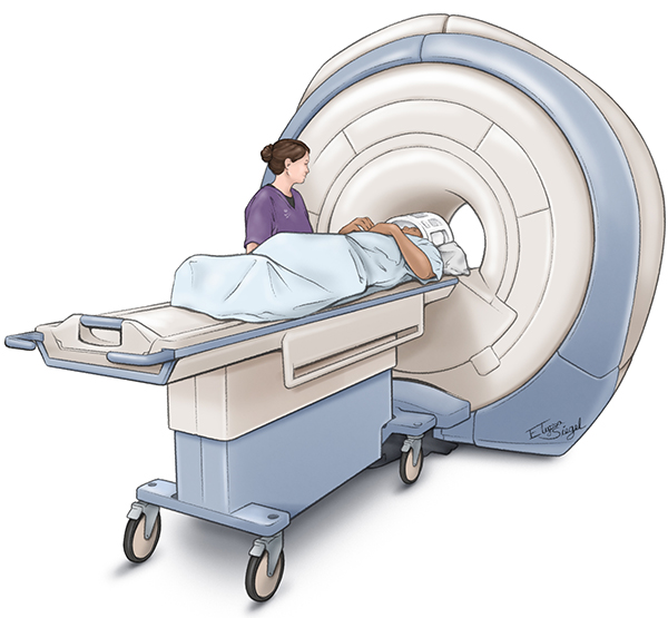
You will be asked to lie very still on the MRI table that is guided into a large magnet. While the procedure is painless, expect to hear various loud, repetitive, banging noises. If desired, earplugs or headphones with music are available to help muffle the noise. Some patients also feel uncomfortable inside the confined space of the MRI scanner. If you have trouble with claustrophobia, talk with your doctor beforehand. An MRI usually takes about 30 minutes but some exams may take longer. If the test is done with contrast, an agent called gadolinium will be administered into a vein in your arm using a needle or small tube.
Less Common Neurosurgical Imaging Tests
Myelogram (Myelography)
A myelogram is an imaging test that uses a contrast dye along with X-rays and/or computed tomography (CT) to examine the inside of the spinal canal, including the spinal cord and its nerve roots. It is used to diagnose conditions such as herniated discs, tumors, bone spurs and infection.
What to expect
With a myelogram, a special contrast dye is carefully injected into your lower spinal canal through a small, hollow needle that is placed under X-ray guidance. The area of the injection is numbed with local anesthetic to limit the pain. The X-ray table will tilt up and down to make the dye flow through your spinal canal. X-rays are taken to record the images formed by the dye. Most often, this is followed by a CT scan that gives even greater detail to the imaging of the spinal cord, nerves, discs and bones.
While you may feel discomfort during the procedure, your healthcare team will take steps to keep you as comfortable as possible. You will need to lie still for a few hours after the procedure.
Angiogram (Angiography)
An angiogram is an imaging test that looks at the blood vessels in the body. Your neurosurgeon will usually order an angiogram test to look for injury or disease involving the blood vessels in your neck, brain or spinal cord. The test is also used to look for aneurysms, blood vessel malformations, some types of tumors and the causes for stroke or hemorrhage.
There are three types of angiogram studies. Computers can use either an MRI scan (MR angiogram) or a CT scan (CT angiogram) to construct images of your blood vessels. The MR angiogram may or may not require the injection of contrast (gadolinium), depending on exactly what issue your doctor is trying to study. A CT angiogram always requires the use of contrast dye.
A formal angiogram is a distinct X-ray test. Our interventional neuroradiology physicians at Goodman Campbell will discuss the angiogram with you. They are specially trained to place a catheter into one of your blood vessels, usually an artery in your groin (femoral artery). Under X-ray guidance, they direct the very small catheter into specific vessels supplying blood to your brain or spinal cord. Contrast dye is then injected through the catheter, and a movie-like series of X-rays shows the blood vessels and how blood is flowing through them.
What to expect
You may be instructed not to eat or drink before any of these angiogram tests. All of these tests require you to lie still while the images are being taken. The MR angiogram is often done during an MRI scan. During a CT angiogram, you will feel a warm sensation for a very brief period of time while the contrast dye travels through your body. A CT angiogram usually takes only a few minutes. The formal angiogram usually is done with multiple sets of pictures being taken. You will feel warmth briefly in the area supplied by the blood vessel that is being studied. You will be watched for several hours following the formal angiogram study to make sure the area where the catheter was placed into the blood vessel has healed well.
PET Scan (Positron Emission Tomography)
A positron emission tomography (PET) scan uses a special camera to record uptake of a radioactive substance (known as a tracer) in different parts of your body. A computer then produces images. A PET scan allows your doctor to look at the metabolic activity of certain tissues or areas in your body to see how they are functioning on a cellular level. PET scans are often done along with a CT scan to get more detailed information.
What to expect
Before the PET scan, a radioactive drug (tracer) is usually given through an IV needle. You must wait until the tracer travels through your body and is absorbed by the organs and tissues before the scan is done.
After a period of time, you will then lie on a table that slides through the PET scanner. The scanner makes a picture of where the tracer is being taken up in your body. For example, cancer cells may show up brighter on the picture than normal, healthy cells. A PET scan can take anywhere from 30 minutes to three hours.
Ultrasound (Ultrasonography)
Ultrasound uses sound waves to make pictures of the inside of your body. Neurosurgeons will use a Doppler ultrasound to look at the blood flow in the carotid arteries in your neck or other arteries in your limbs. Ultrasound is also used to look at various organs or abnormal areas such as tumors, cysts, abscesses or blood clots. Ultrasound does not use any X-rays. High-frequency sound waves are sent from a small probe through a gel placed on your skin. The sound waves sent back are collected by the probe and a computer uses the sound waves to create an image.
What to expect
Ultrasound gel and a small probe (transducer) are placed directly on the skin over the area to be studied. Ultrasound is not painful, but you may feel some pressure.
Request an appointment online and we’ll guide you through the next steps.