What Is Thoracic Spinal Stenosis?
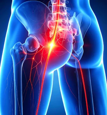
Thoracic spinal stenosis is a condition in which the spinal canal located in the thoracic spine — the middle portion of your back — becomes narrowed, placing pressure on the spinal cord. The thoracic spine is made up of 12 vertebrae, each connected to the ribcage, creating a stable yet flexible structure that supports the upper body and safeguards the spinal cord. This region plays a vital role in posture, movement, and overall spinal health.
While cervical stenosis occurs in the neck and lumbar stenosis in the lower back, thoracic spinal stenosis is less common but can be more serious due to the increased likelihood of spinal cord compression in this area. Compression in the thoracic spine can lead to significant pain, neurological symptoms, and, in severe cases, loss of mobility or bladder and bowel control.
Patients experiencing symptoms or looking to prevent progression may benefit from specialized spinal stenosis treatment in Indiana, where access to advanced diagnostics, skilled spine specialists, and comprehensive care can make a substantial difference. Early detection and timely expert intervention not only help relieve discomfort but also reduce the risk of long-term complications, ultimately supporting a better quality of life.
What Causes Thoracic Spinal Stenosis?
There are numerous potential thoracic spinal stenosis causes, ranging from age-related changes to conditions present at birth. One of the most common is degenerative spine disease, a gradual process in which wear and tear over time leads to changes such as arthritis in the spinal joints, thickening of ligaments that normally provide stability, or deterioration of the intervertebral discs that act as cushions between the vertebrae. These changes can progressively narrow the spinal canal, placing increased pressure on the spinal cord.
Trauma is another significant factor. Serious falls, motor vehicle accidents, or high-impact sports injuries can fracture or displace vertebrae, damage discs, or cause swelling that leads to stenosis. In certain cases, abnormal growths such as tumors develop within or around the spinal canal, creating direct compression of the spinal cord and disrupting normal nerve function.
Some individuals are born with congenital spinal stenosis, meaning their spinal canal is naturally narrower than average; while symptoms may not appear immediately, this structural difference can predispose them to more severe issues later in life. Other contributing factors may include chronic inflammation from autoimmune conditions, vascular abnormalities that alter blood flow around the spine, or complications, such as scar tissue formation, arising from previous spinal surgeries.
What Are the Symptoms of Thoracic Spinal Stenosis?
The most common thoracic spinal stenosis symptoms often begin subtly but can become increasingly disruptive over time. Persistent pain in the mid-back is frequently reported, sometimes accompanied by discomfort that radiates around the ribs or wraps across the chest in a band-like pattern. Because the thoracic spine protects and surrounds the spinal cord, narrowing in this region can lead to significant neurological symptoms, including numbness, tingling sensations, or muscle weakness in one or both legs. These changes may be mild at first, such as occasional leg heaviness or fatigue, but can progress to noticeable difficulty climbing stairs, standing for long periods, or maintaining a steady walking pace.
Many patients also develop changes in balance or coordination, which can increase the risk of falls and make daily activities more challenging. In more severe cases, spinal cord compression can disrupt nerve signals to the bladder or bowel, causing incontinence or difficulty controlling these functions — symptoms that require urgent medical attention. Because these signs can sometimes resemble those of other conditions, from musculoskeletal issues to neurological disorders, seeking evaluation from a qualified spine specialist is essential for accurate diagnosis. Left untreated, thoracic spinal stenosis can continue to progress, leading to more pronounced symptoms and potentially permanent nerve damage, underscoring the importance of early recognition and intervention.
How Is Thoracic Spinal Stenosis Diagnosed?
Thoracic spinal stenosis diagnosis typically begins with a detailed medical history, where the healthcare provider reviews the patient’s symptoms, their duration, any prior injuries or surgeries, and underlying health conditions that could contribute to spinal problems. This is followed by a comprehensive physical examination, during which the clinician evaluates posture, range of motion, and areas of tenderness or muscle spasm. A focused neurological exam is a critical part of the process, assessing muscle strength, reflex responses, coordination, and sensation in the legs to determine whether spinal cord or nerve function is impaired.
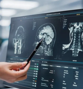
Imaging tests are often used to confirm the diagnosis and pinpoint the location and severity of narrowing. An MRI is considered the gold standard for visualizing the spinal cord, intervertebral discs, ligaments, and other soft tissues in high detail. In some cases, a CT scan is used to obtain cross-sectional images that reveal subtle bony changes, fractures, or structural deformities, while standard X-rays can show spinal alignment, vertebral damage, or degenerative changes.
When necessary, additional advanced studies — such as myelography, electromyography (EMG), or nerve conduction studies — may be performed to evaluate nerve function or rule out conditions that mimic thoracic spinal stenosis. Collaborating with a team highly experienced in thoracic spinal stenosis diagnosis ensures that each step of the evaluation is precise, allowing for a clear understanding of the underlying problem and the creation of a tailored treatment plan that addresses the patient’s specific needs.
What Are the Treatment Options for Thoracic Spinal Stenosis?
The right approach to managing thoracic spinal stenosis depends on multiple factors, including the severity of the condition, the underlying cause of the narrowing, the degree of spinal cord compression, and the patient’s overall health and activity levels. In many cases, physicians will consider nonsurgical treatment aimed at relieving symptoms, improving mobility, and preventing further progression. These may include physical therapy programs designed to strengthen core and back muscles, enhance flexibility, and improve posture to reduce strain on the spine. Medications may also be prescribed to help manage pain and reduce inflammation, while targeted lifestyle modifications, such as weight management, activity pacing, and ergonomic adjustments, can ease daily strain on the thoracic region.
If conservative measures fail to provide relief or if neurological symptoms worsen, surgical treatment may become necessary to prevent permanent nerve damage. Common surgical procedures include a laminectomy, in which part of the vertebra is removed to create more space for the spinal cord, and spinal fusion, which stabilizes the spine by permanently connecting two or more vertebrae. In cases where a tumor, lesion, or other abnormal growth is causing compression, surgical removal may be required as part of the treatment plan. Advances in minimally invasive techniques now allow many patients to benefit from smaller incisions, reduced muscle disruption, shorter hospital stays, and faster overall recovery compared to traditional open surgery.
For the best possible outcome, thoracic spinal stenosis treatment should be coordinated by an expert, multidisciplinary spine care team like that at Goodman Campbell. This approach ensures that each aspect of the patient’s condition — from diagnosis to rehabilitation — is addressed with specialized knowledge, collaborative planning, and a personalized strategy that supports long-term spine health and quality of life.
When Should You See a Doctor for Thoracic Spinal Stenosis?
It is important to understand when to see a doctor if you begin to experience symptoms that could indicate serious spinal cord involvement. Warning signs such as sudden weakness or numbness in one or both legs, noticeable changes in balance or coordination, or a loss of bladder or bowel control should never be ignored. These symptoms may signal significant spinal cord compression and require urgent medical attention to prevent further damage. Delaying seeking treatment not only increases the risk of permanent nerve injury but can also make recovery more challenging. A qualified spine specialist has the expertise to conduct a thorough evaluation, determine the underlying cause, and guide you through the most effective treatment options, whether they involve nonsurgical interventions or advanced surgical procedures.
For individuals in Indiana, scheduling a Goodman Campbell consultation offers the opportunity to connect with a multidisciplinary team of spine experts who can provide comprehensive care, personalized recommendations, and ongoing support on the path to recovery. Early action is often the key to preserving mobility, protecting nerve function, and maintaining quality of life.
How Can Goodman Campbell Help?
Goodman Campbell offers specialized spine care through a team of experts who work together to diagnose and treat complex spinal conditions. Their approach begins with a comprehensive evaluation to pinpoint the exact cause of symptoms, followed by the creation of a personalized treatment plan. Patients have access to both nonsurgical therapies and advanced surgical options, including laminectomy and spinal fusion. Equally important, Goodman Campbell provides ongoing support throughout the recovery process, ensuring patients are informed, supported, and confident at every stage. If you are ready to take the next step, you can request an appointment directly or ask your physician for a referral.
Thoracic spinal stenosis is a serious condition that can significantly impact daily life, but early detection and proper treatment can lead to better outcomes. Recognizing causes of thoracic spinal stenosis, paying attention to symptoms, and obtaining an accurate diagnosis are essential steps in protecting your mobility and quality of life. If you or someone you know is experiencing signs of this condition, do not delay — seek treatment from trusted specialists, such as the team at Goodman Campbell, to ensure you receive expert care tailored to your needs.